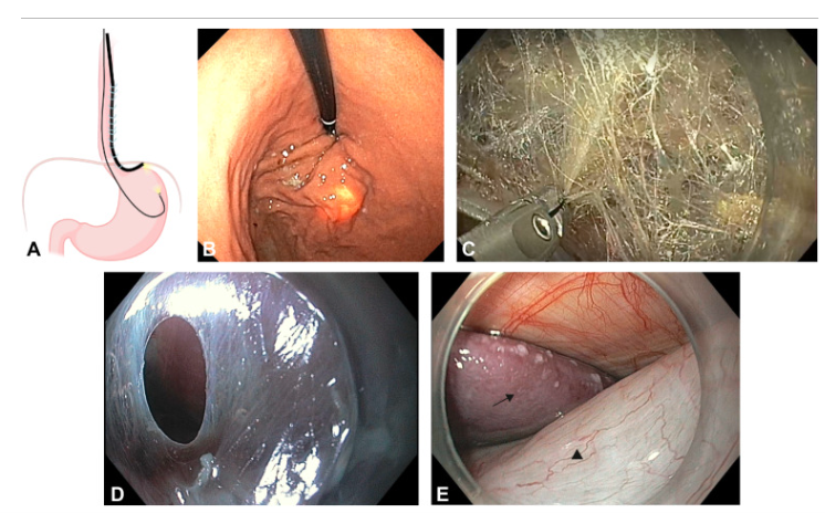
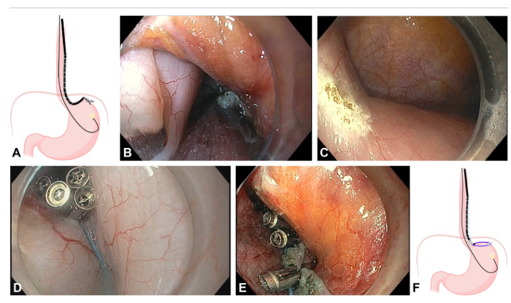
Anchoring the Loop Ligating Device
A, Schematic representation of the wrap simulation by grasping the anterior wall of the gastric fundus and retracting while under visualization by the ultraslim gastroscope. B, The fundic serosa was pulled to the edge of the gastric myotomy using a grasping forceps. C, An appropriate point on the fundic serosa was marked using cautery. D, The loop ligating device fixed at the fundic serosa (distal anchoring point) using clips. E, Attachment of the loop ligating device at the distal end of the right edge of the myotomy (proximal anchoring site) with clips. F, Schematic drawing of the loop ligating device attached at the proximal and distal anchoring sites.