Researcher Developing Microscope that Can Shorten Time from Biopsy to Cancer Diagnosis |
|
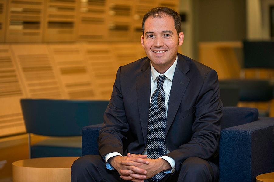
J. Quincy Brown, PhD, and his team are developing a microscope that can examine biopsied tissues in the operating suite, with a goal of speeding diagnoses and moving toward a 'see-and-treat' paradigm for prostate and other cancers. |
|
"Many men have unfortunately had the experience of getting a prostate biopsy and then waiting a week or two for the results, and only then can their physician determine whether they have cancer and if so, how to treat it," said J. Quincy Brown, PhD, associate professor of Biomedical Engineering.
Shortening the time from biopsy to diagnosis can have many positive results. It can reduce the anxiety the patient and his or her family must endure while they wait for answers, but it can also mean they start their treatment plans sooner, which is extremely important to positive outcomes.
Brown and his team are addressing this issue by creating microscopes that can quickly and accurately provide pathology at the point of care. "If we can start moving the ability to look at the tissue microscopically closer to the patient in space and in time — meaning right into the operating room — then we can move toward a future 'see-and-treat' paradigm for localized prostate and other cancers," said Brown.
The field is called ex vivo microscopy, and Brown and his team are at the center of it, having invented and patented a microscope that can image fresh bulk tissue in the operating room within minutes of removal, without the need for histological staining or slicing required by current pathology methods.
"The reason histopathology takes so long is that the tissue must be stained and sliced into microscopically thin sections in order to pass light through it," said Brown. His team's new microscopes don't require light to pass through the tissue. They can isolate images from certain planes of the tissue that they virtually cut with light, providing a clear picture of the surface. The images look like traditional histological images, but they're not. They're captured in minutes and in a totally different way.
"What if a patient who sees their physician for a prostate biopsy could have been a candidate for a localized ablation but we lose them to follow-up while waiting for the pathology results," said Brown. "If you're already doing the biopsy procedure, wouldn't it be better if you could image the tissue in real time, have a pathologist standing by to analyze those images to determine if the tissue is cancerous and then map where the tumor is with a goal of ablating — or removing — the tumor right then and there?"
Localized ablation is an emerging treatment for prostate cancer that minimizes recovery time and preserves healthy function in eligible patients. However, it currently takes a week or two following a biopsy to determine if the patient is a candidate for it.
"We utilized tissue samples from 39 patients undergoing diagnostic biopsy at Tulane Medical Center," said Brown. "We asked urologists to give their best guesses using the imaging tools they have now — MRI and ultrasound — if they were going to do a see-and-treat, would they do it on each of the patients biopsied. A comparison of the urologists' suspicions of malignancy to pathologist diagnosis based on biopsy images obtained by our microscope reveals that real-time biopsy imaging could significantly improve confirmation of malignancy over medical imaging alone."
Brown's team is utilizing the same technology to intra-operatively image and examine the surface of surgically removed prostate glands to determine whether margins are clear. "We've developed an automated prostate positioning system that lifts, lowers and rotates the gland while the microscope takes panoramas — thousands of images that are then stitched together to give us a map of the surface of the tissue. If there is any tumor on the surface of the prostate, then by extension you would assume there is also cancer remaining in the patient, and so you could go back to that associated location and take more tissue while the patient is still in the OR."
"The utilization of real-time assessment of the location of prostate cancer could revolutionize therapy for the more than 200,000 men diagnosed with the disease each year," said L. Spencer Krane, MD, Chief or Urology for the U.S. Department of Veterans Affairs, and collaborator on this project. "Understanding where the cancer is, and importantly where the cancer isn't, with this level of specificity could allow surgeons to preserve the vital structures adjacent to the cancer, while still ensuring complete eradication of the disease. This is a game-changing technology when applied to prostate cancer patients."
Next steps are to make both processes even faster and move them toward clinical trials to see if they improve outcomes.
Brown stresses the importance of the interdisciplinary nature of this work. "I really believe in the potential of team science, with engineers and physicians working closely together," he says. In addition to Krane, Dr. Shams Halat, associate professor of pathology, collaborated on this project.
"I look forward to continuing our collaboration with Brown's team, to move this technology from the bench to the bedside in the coming years," said Krane.
|
|
Madeline Behr
Biomedical Engineering
PhD Candidate
|
|
Shams Halat, MD
Associate Professor of
Pathology
|
|
L. Spencer Krane, MD
Chief of Urology,
U.S. Department of
Veterans Affairs
|
|
Robotic Bronchoscopy Program Helping Detect Lung Cancer Earlier Than Ever Before |
|
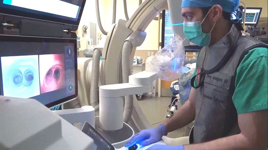
Dr. Ramsy Abdelghani utilizes real-time cone beam CT scan imaging to help guide a robotic bronchoscope directly to a small nodule in a patient's lung. |
|
That's how he describes the capabilities of Tulane's state-of-the-art Robotic Bronchoscopy Program, which combines a robotic bronchoscope with real-time advanced cone beam CT scan imaging, allowing him to precisely navigate to virtually anywhere within the lungs and biopsy smaller lesions than ever before. This leads to earlier diagnoses of lung cancer, essential for good outcomes. Tulane has performed more of these advanced procedures than any other program in the state.
The robot utilizes a pre-procedural CT scan to precisely isolate the location of the nodule and create a suggested route for the bronchoscope — GPS-like directions that allow Dr. Abdelghani to guide a thin, 360-degree maneuverable catheter through the numerous airways that make up the lungs directly to his target. And some of these nodules are as small as 3.5 mm in diameter — approximately three times smaller than those he could biopsy previously.
"I use a console to drive the robot, almost like a video game," said Abdelghani. "Without guidance, this would be extremely challenging. These airways all look the same and there are hundreds of wrong turns I could make along the way."
Once the target is reached, a low-dose CT scan allows Dr. Abdelghani to see the spatial relationship of the catheter to the nodule, make adjustments if necessary and then biopsy with confidence that he's in the correct position to retrieve a good tissue sample. "If the pathology comes back benign, I can say with relative certainty that I got a good representation of the lesion," said Abdelghani. "The way I think about it is the robot gets me to the driveway, but the cone beam CT scan gets me to the front door."
All Tulane Medical Center patients with lung nodules are now referred to Dr. Abdelghani's clinic. Previously, small lung nodules that incidentally showed up on imaging tests of the heart or abdomen may not have been candidates for biopsy because they were too small. "But since the availability of robotic bronchoscopy, I haven't said no yet," said Abdelghani, who has biopsied approximately 250 nodules in the year since the program started.
Robotic bronchoscopy requires general anesthesia. However, the potential risks of this scarless, well-tolerated, virtually pain-free procedure are quite low, and Dr. Abdelghani says most patients go home the same day.
If you are interested in learning more or making an appointment with one of Tulane's board-certified interventional pulmonologists, please contact Dr. Abdelghani at rabdelgh@tulane.edu or call Tulane's Lung Nodule Clinic at 504-988-8600. |
|
Findings Show Substantial Racial, Ethnic and Geographic Disparities in Triple Negative Breast Cancer Rates |
|
A new study by researchers at the American Cancer Society (ACS) and the University of Texas Health Science Center shows substantial racial and geographic variations in incidence rates of triple-negative breast cancer (TNBC) among women in the United States. The study found Black women in Delaware, Missouri, Louisiana, and Mississippi have the highest TNBC incidence rate, more than four times as much, compared to Asian or Pacific Islander women with the lowest rate. The study is published in the Journal of the American Medical Association (JAMA) Oncology.
"What we found is that even within racial and ethnic groups, variation based on a women’s state of residence was considerable,” said Dr. Hyuna Sung, lead author of the study and senior principal scientist at the American Cancer Society. “Within each racial and ethnic group, there were substantial differences in incidence rates across state lines. Intriguingly, for both Black and white women, rates were lowest in Utah and highest in Iowa, Mississippi, and West Virginia among the states with data available for both Black and white women. This data suggests that social, environmental, and structural determinants of health are at play in shaping the geographically patterned risk of TNBC and this merits further study.”
TNBC accounts for 10% to 20% of all breast cancer diagnoses and is called “triple negative” because the cancer cells do not have progesterone or estrogen receptors and do not make any HER2 protein – the cells test negative for the biomarkers of these receptors or proteins. Breastfeeding is the one established protective factor for this otherwise aggressive form of breast cancer. Evidence also suggests that increased physical activity and high levels of fruit and vegetable consumption may reduce the risk of TNBC, whereas alcohol consumption and premenopausal obesity may increase the risk of TNBC.
For the study, the team of researchers used population-based cancer registry information from the U.S. Cancer Statistics Public Use Database. This included statistics from all 50 states and Washington, D.C. from January 2015 through December 2019. Racial and ethnic information was included based on self-reported or inferred data from medical records and classified into mutually exclusive categories of Hispanic, non-Hispanic American Indian or Alaska Native, non-Hispanic Asian or Pacific Islander, non-Hispanic Black, or non-Hispanic white. Age-standardized incidence rate of TNBC was calculated by race and ethnicity, then incidence rate ratios were calculated to quantify disparities in state-specific rates.
Results from the study showed that non-Hispanic Black women in Delaware, Missouri, Louisiana, and Mississippi were diagnosed with TNBC at the highest rate of 29 per 100,000 women per year. Asian or Pacific Islander women in Oregon and Pennsylvania saw the lowest rate of less than seven per 100,000 women per year. Black women in Delaware, Missouri, Louisiana, and Mississippi experienced a rate of diagnosis that was more than double what white women in those same states experienced.
“It is important to identify and support states where the benefit of breast cancer prevention and surveillance efforts have the greatest impact,” said Sung. “We should also promote and fund breast cancer research that has a broad representation of racially and ethnically diverse populations to better understand the roles of social determinants of health in racial and geographic disparities in the burden of TNBC.”
Resources from the American Cancer Society on breast cancer risks, screening, and treatment can be found here.
|
|
Will You Go to the Prom With Us?
And Help Raise Funds for Breast Cancer Research
|
|
Want a chance to re-live (or reinvent!) your prom night while honoring breast cancer survivors?
Join us for the Krewe de Pink Prom, a fun-filled evening organized by Krewe de Pink, a local, all-volunteer nonprofit that raises funds to support breast cancer research at Tulane Cancer Center.
This year's event takes place on Saturday, September 30, 2023, 7 - 11 PM, at Generations Hall, 310 Andrew Higgins Blvd.
Come dressed in pink prom style, take a prom photo and then dance the night away to your favorite disco, funk, jazz, Motown, R&B and salsa tunes, courtesy of New Orleans’ own Crescent City Soul. You might event get crowned Prom King or Queen!
Other entertainment includes a special performance by the Pussyfooters, and breast cancer survivors in attendance will be honored and celebrated with a dedicated "promenade."
This 21 & older event includes a cash bar, featuring the winning entries from Krewe de Pink’s 2023 Pink Cocktail Contest. Attendees may also bring along snacks for their table.
The auction features a variety of items, including New Orleans Staycation packages, jewelry, artwork and one-of-a-kind, hand-decorated costume bras. There's also a raffle, featuring a wine barrel, a beer bucket, and a Lotto ticket bundle! You can view and bid on select auction items NOW by clicking here, even if you're not able to attend the event .
|
|
or visit
To make a tax-deductible donation to Tulane Cancer Center
(Federal Tax ID# 72-0423889) please click here.
Thank you for your generosity and support!
|
|
|
|
|
|
|