| |

Tech
Update
|
Neuroscience Edition, November 2017
|
Welcome to Scitech's Neuroscience newsletter.
The main focus of this newsletter is to introduce new products and technologies relevant to Neuroscience Research. Scitech will be exhibiting at ANS 2017 at the International Convention Centre, Sydney from 3-6 Dec 2017. Please visit Scitech at Booth 11 to discuss and see some of the latest technologies designed to help facilitate your neuroscience research.
|
|

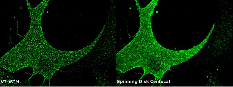
High Speed Super Resolution Imaging System
Visitech: VT-iSIM
The VT-iSIM high speed super resolution imaging system from VisiTech International, produces super resolution images in real time at up to 1,000 frames per second. The VT-iSIM can image at spatial and temporal resolutions and does not require any specific fluorophores, immersion oils, objective lenses or even microscope frames to work. If you have a sample which fluoresces you can enhance your spatial and axial resolution by up to 2X regular for wide-field microscopy.
Features:
- Super Resolution Imaging
- X,Y <125nm, Z<350nm
- Multi Point Scanning Technique
- Low Photo Bleaching
- Fast Scanning (>1000fps)
- Perfect Camera Sync
|

Quantitative Imaging Software for Stereology, Neuron Reconstruction and Image Analysis
MBF Bioscience: Neurolucida version 2017
With the latest Neurolucida 2017, neuroscience researchers can reconstruct and analyze neurons with greater speed, efficiency and ease.
Neurolucida 2017 is the system of choice for neuron tracing and reconstruction, neuron analysis and 3D brain mapping. It integrates with the latest microscopic imaging devices and computer hardware, enabling you to trace neurons with cutting-edge microscope technology.
New Features
- Edit reconstructions in an interactive 3D environment when tracing image stacks
- Improved handling of big image data
- New analyses in a redesigned Neurolucida Explorer- the comparison software to Neurolucida which performs all of the quantitative analyses generated from neurons is digitally reconstructed with Neurolucida
Contact Us
MBF Bioscience: Stereo Investigator 2017
The brand new Stereo Investigator 2017 package enables users to collect unbiased stereology data with greater speed and effectiveness. It provides users with cutting edge technology to break down the characteristics found within tissue specimens, including the nature of cells and biological structures. It integrates the latest microscopic imaging devices and computer hardware.
New performance features include
- Edit contours and markers in an interactive 3D environment
- Improves handling of big image data
- An entire ribbon bar dedicated to publishing results
|

Multi Channel In-Vivo Electrophysiological Recording
Blackrock Microsystems: CerePlex™ Direct
The CerePlex™ Direct is a sophisticated direct digital recording data acquisition system, providing unprecedented low-noise recording in a simple plug-and-play package. CerePlex™ Direct is ideal for small mammalian models, non-intensive electrophysiologists, and beginners in data acquisition recording and stimulating.
The system is designed for use with any of Blackrock's CerePlex™ digital head stages and is also compatible with the NeuroMotive™ System for video tracking.
Blackrock Microsystems: 1gram Digital Headstage
The
CerePlex™ µ is a very small and lightweight (1 gram) digital headstage that provides a platform for simple stimulation and tracking in freely moving rodents. It
provides an interface between the Cerebus™ or CerePlex™ Direct recording systems and up to 32 microelectrodes for high-fidelity transmission and recording of extracellular spikes and local field potentials from the brain.
In addition to neural recording, it features an onboard 3 axis magnetometer, accelerometer, and gyrometer as new tools for monitoring animal behavior.
Learn More
|
The Future of Scientific sCMOS Camera technology is here now!
Photometrics sets the new standard in scientific sCMOS camera technology
Photometrics: Cameras suitable for Low Light Fluorescence microscopy and Light Sheet microscopy
- Prime 95B Camera
Back Illuminated, 95% QE, 1.2 MP, 11 um pixels, -10 deg C cooling, PCIe and USB3.0
- Prime BSI Camera
(New)
Back Illuminated, 95% QE, 4.2MP, 6.5um pixels, -10 deg C cooling,
PCIe and USB3.0
- Prime 4.2 Camera
72% QE, 4.2 MP, 6.5um pixels,-10 deg C cooling, PCIe and USB3.0
Learn More
Contact Us
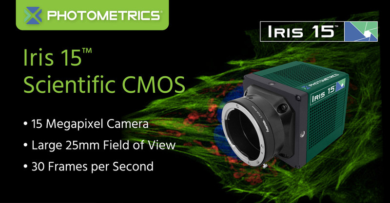
Photometrics: Cameras suitable for Large Field of View imaging and Light Sheet microscopy
IRIS 15 Camera (New)
73% QE, 15 MP, 4.25 um pixels,-10 deg C cooling, PCIe and USB3.0
IRIS 9 Camera (New)
73% QE, 9 MP, 4.25 um pixels,-10 deg C cooling, PCIe and USB3.0
|

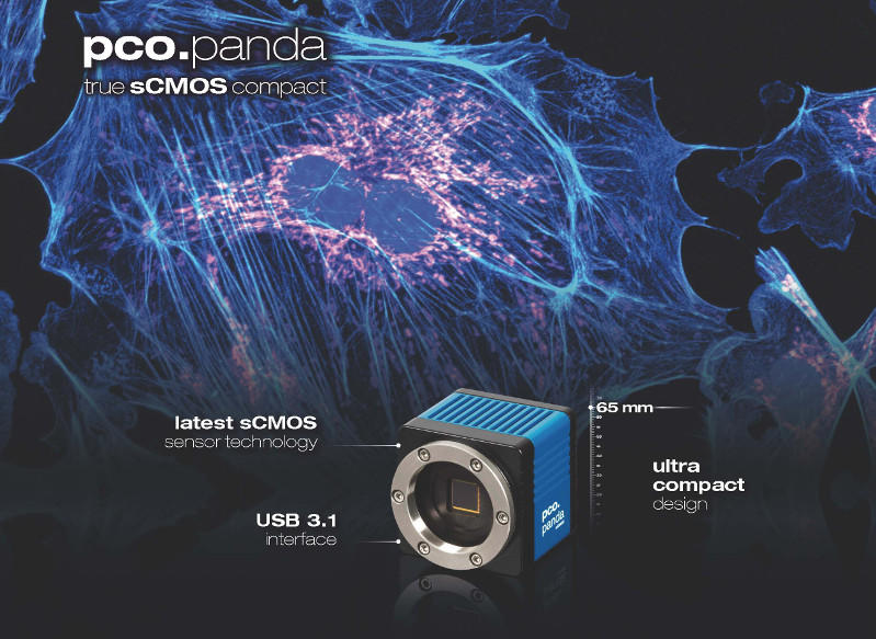
PCO: pco Panda - Smallest 4.2MP sCMOS Camera
The pco.Panda sCMOS camera is ideal for applications such as GSDIM, PALM, STORM, SPIM, SIM, live cell microscopy, single molecule detection, light-sheet microscopy, spinning disk confocal microscopy, FRET, FRAP, fluorescence spectroscopy, bio- and chemi- luminescence and high content screening. It includes the latest 16-bit sCMOS sensor technology, dynamic range of 22,500:1, exposure time from 100 µs to 1s, maximum frame rate @ full resolution > 40 fps and rolling shutter.
Key Features include:
- QE up to 80%
- low readout noise of < 2 e
- high resolution of 2048 x 2048 pixels
- pixel size of 6.5 μm
- Compact body measures 65 x 65 x 65 mm³
- Weighs as little as 450g (without lens adapter).
- USB 3.1 interface
|

Electrophysiology &
Multi-Photon Microscopy Systems
Scientifica: VivoScope
Scientifica specializes in the development, design and manufacture of high quality solutions for applications including patch clamp and extracellular electrophysiology, two-photon imaging and optogenetics studies.
Scientifica's Vivoscope is an industry leading in-vivo imaging system for two-photon behavioural studies. It is ideal for in vivo samples, linear or spherical treadmills, large stereotaxic frames or other virtual reality setups. It integrates acoustic insulating foam to dramatically reduce audible noise, and provides deep imaging for large populations of cells. The fast frame rate is instrumental in helping users accurately position the patch pipette under the infrared light. Glavo or Resonant scan head options are available, depending on the nature of experiment that is being undertaken.
Benefits include:
- Galvo or Resonant scan head options available
- Integrates acoustic insulating foam to dramatically reduce audible noise
- Deep imaging for large populations of cells
- Patch under multi photon illumination
- Extra space between light path and sample

Contact Us
|
M
ultielectrode arrays (MEA) for high resolution In-vitro extracellular electrophysiology

3Brain: BioCam X
The BioCam X from 3Brain is a large-scale sensing Microelectrode Array platform for high resolution, label-free in-vitro electrophysiological imaging. It allows simultaneous recordings from a total of 4096 electrodes sampled at 18 kHz per electrode thus allowing monitoring of thousands of neurons. At the core of the MEA Biochip is an active monolithic CMOS device.
Applications include:
- Human stem cell-derived neurons
- Acute brain slices
- Drug discovery
- Plasticity studies,
- Cardiomyocyte cultures and
- Ontogenetic-combined studies
|

Deep Brain Imaging & Stimulation Systems for Freely Behaving Animals
Mightex Systems: OASIS Implant
The OASIS Implant from Mightex Systems is a ground-breaking micro-endoscopic system that is capable of doing deep-brain simultaneous all-optical recording and manipulation of neural circuit activity with sub-cellular resolution, on freely behaving animals. Compatible with high-sensitivity research-grade cameras, the OASIS Implant enables researchers to acquire high quality deep-brain images & videos. In addition, with multiple illumination ports, OASIS Implant is equipped with both wide-field and patterned illumination capabilities (when combined with Mightex's market-leading Polygon400) and it is also capable of doing multi-wavelength imaging/stimulation which cannot be done with the existing head-mounted cameras. The overall weight of the head-mounted fixture is as little as 0.3g making OASIS Implant an ideal research tool for in-vivo live animal experiments.
Other applications include:-
- Deep brain calcium imaging in freely behaving animals
- Deep brain patterned optogenetic stimulation with single-cell or sub-cellular resolution
- Simultaneous deep brain stimulation and imaging
Learn More
|

Ultrafast Laser System for Multiphoton Microscopy
Chromacity Lasers: Spark 1040
The Spark 1040 Laser System is a cost effective, air-cooled, compact, ultra-fast ytterbium fibre-based laser providing exceptional performance with turnkey operation and is available in two versions, delivering either 500 mW or up to 2.5 W, with picosecond or femtosecond pulse-widths and operating at a wavelength of 1040 nm.
Applications include:
- Multiphoton microscopy
- Light Sheet microscopy
- Optogenetics imaging experiments
- Fluorescence measurement of dyes/quantum dots
- Time resolved experiments (TCSPC/FLIM)
In Multiphoton Optogenetics experiments, it has 3 key benefits:-
- Inherent 3D resolution allows cells/groups of cells to be imaged at typically micron resolution in the z axis and a few hundred nm in the XY plane. Using a pulse laser source also allows time resolved measurements to be taken
- Images can be captured at greater sample depths by the use of 2x or 3x longer wavelengths resulting in a 16x or 81x reduction in scatter as well as an increase in image depth
- Photobleaching and photothermal degradation is reduced, allowing in vivo experiments to be more readily accessible

|
Melb: +61 3 9480 4999
Syd: +61 2 9705 8059
Australia Wide: 1300 724 832 ( 1300 SCITECH )
|
|
|
|
| |
|